
Simulation
Introduction
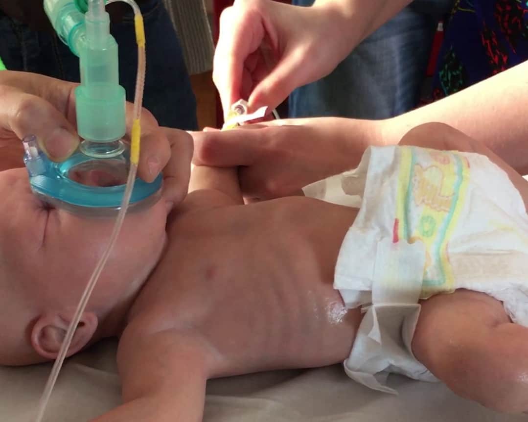
The course concludes with candidates undertaking three simulated scenarios. These scenarios provide candidates with an opportunity to consolidate their learning by drawing upon theory and skills learned during the course. Simulation based medical education enables learning in a realistic and safe environment. This chapter briefly discusses the history of simulation in endotracheal intubation. In addition each of the three scenarios are discussed and the key learning points from each are identified.
History of simulation and endotracheal intubation
The eighteenth century saw severe epidemics of diphtheria in France, the United Kingdom and North America. The first symptoms of diphtheria include malaise, throat pain and mild fever. After 2-3 days a thick layer of dead mucosal cell debris, a pseudomembrane, develops in the pharynx, larynx and trachea. The pseudomembrane is a result of toxin released from the causal bacteria. This was a major cause of death in children who were at particular risk of airway obstruction and subsequent death from suffocation because of their narrow airways. As a result there was a widespread drive to develop clinician’s skills in airway management.1
1. Cadavers
Between 1880 and 1885 Joseph P O’Dwyer, a paediatrician in New York, experimented on cadavers and patients in an attempt to develop a system of intubation for airway obstruction in diphtheria.2 In 1887 he demonstrated his method of intubation on a cadaver at the annual meeting of the Medical Society of the State of New York. 3 Later that year Jasper Garmany published ‘Operative Surgery on the Cadaver’ in which he described how to perform a wide range of skills, including tracheal intubation.4 Soon practicing intubation on cadavers had become common place. Many practitioners commented on the importance of practicing on cadavers:
‘I do not think anyone should attempt the operation without previous practice on the cadaver. Without this practice failure is almost certain to result.’ G.McNaughton5
‘To avoid accidents, it is very essential to have some preliminary practice on the cadaver’ F.Huber6
In some instances, when cadavers were unavailable, dogs were used to practice the sequence of steps required for intubation. However, their anatomy was very different and the benefit of this was questionable.
2. The first intubation simulators
In 1893 Otto Heubner, a paediatrician working in Germany, described the limitations of using cadavers to gain expertise in intubation. He highlighted that they were not always readily available. In addition the body couldn’t be used after rigor mortis had developed and that the loss of muscle tone after death meant the relationships between anatomical structures were not necessarily similar to those in a live patient. In response he created the first larynx simulator for learning intubation. To make the simulator a cavity in a shape that corresponded to the open mouth and bony supports of the pharynx and larynx was cut in a large block of cork. The tongue, throat parts, larynx and trachea were then removed from a cadaver. These were then attached to the cork. This model could be used for a period of weeks if kept in an appropriate preserving fluid. The following year Amand Schlossarek demonstrated a simulator for learning intubation at a meeting of the College of Physicians in Vienna. Scholossarek’s phantom had a larynx made from an elastic material mounted in the normal position in the neck of a bust of a child. The model was designed to represent a bust of a choking child in the upright position. It was made by Herr Reiner, an Austrian instrument maker, and an illustration was included in an article for teaching aids for ENT. 7 From the late nineteenth century simulation was widely used to teach intubation in US medical schools.
3. Later use of simulation
The need to teach emergency intubation reduced in the twentieth century with the development of antitoxin and a significant reduction in the risk of death from diphtheria. Vaccinations have now eradicated the disease from most developing countries. However, the importance of oro-tracheal intubation in the management of medical emergencies and trauma was described in the second half of the twentieth century. Consequentially clinicians once again turned to cadavers and simulators to practice. A prototype laryngoscopy simulator with a flexible rubber airway was developed in Canada.7 Over the last 20-30 years there has been a marked increase in the production and availability of airway simulators.
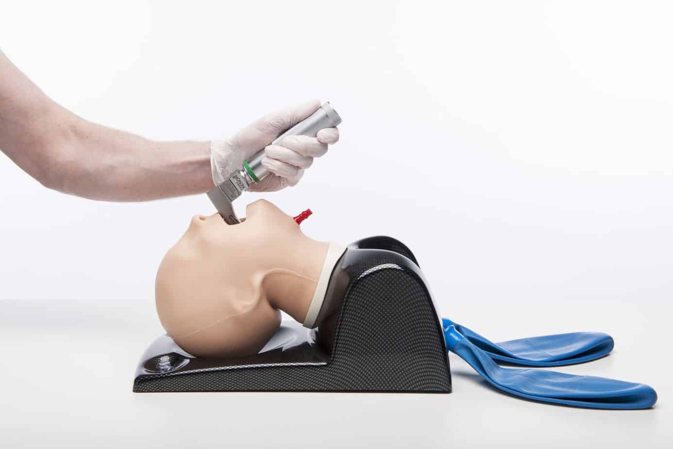
Trucorp AirSim® Child
There has also been a significant improvement in quality both in relation to visual appearance and feel. 4D printing has enabled the creation of highly realistic mannequins. In addition simulation training has evolved to focus not only on the technical skill of intubation but also to emphasise the importance of avoiding multiple attempts and planning for and dealing with difficult airways.
Already attended the course?
If you have already attended the Paediatric Emergencies Intubation Course you can view the learning points from the course simulation scenarios by clicking on the link below. As this section is designed to be viewed only after completing the course you will need to enter the password you were given on the course to access this section.
References
- Owen H. (2016) Simulation and Teaching in Resuscitation and Trauma Management. In: Simulation in Healthcare Education. Springer, Cham
- Sperati G, Felisati D. Bouchut, O’Dwyer and laryngeal intubation in patients with croup. Act Otorhinolaryngol Ital. 2007;27(6):320–3
- O’Dwyer J. Demonstration on the Cadaver of the Method of Intubation of the Larynx, Meeting of the Medical Society of the State of New York on Feb 3rd 1887. Med Record. 1887;31:160
- Garmany J. Operative surgery on the cadaver. New York: D Appleton; 1887.
- McNaughton G. Intubation of the larynx. N Y Med J. 1877;45:624–6
- Huber F. Intubation. Med Record. 1887;31(25):677–84.
- Bennett MR. A model for teaching laryngoscopy. Can Anaesth Soc J. 1954;1(2):123–5
![]()